Saturday, December 25, 2010
At the end of my second week in clinicals 12-23-2010
I spent two weeks without Kim at my clinical site and I have worked with many techs. I am ready to work with my clinical instructor, Kim, and work at perfecting my abdomen scanning routine. Many patients have gas, and it is hard to find the perfect window for the liver and great vesels. I will try to enjoy my week off, and return to the site on January 3rd, after spending some quality time with my family. Merry Christmas and Happy New Year!!!
After one full week of clinicals
I spent one full week at my clinical site. I rotated from the suite 100, to the women center and even to the vascular lab, suite 201. It has been interesting and I am ready to start scanning myself soon.
My first day of clinicals 12-13-2010
Today I started my clinical rotation. I am assigned to SMIL, Desert Mountain office, on Shea and 101. It is a long drive and I left one hour and a half prior to my starting time. I got there on time, and found where to go to sign in on the computer. I met Kim and I was introduced to all the other technologists. I spent my first day with Stephanie Flores, following the techs and observing their routines and preferences.
Thursday, December 2, 2010
DMS 110 lab 12-2-10 Thyroid
After one week off ( Thanksgiving week) we returned to DMS 110 lab.
Today we scanned for the first time the thyroid, both in trans and long. I measured the gland in long. We also used for the first time the dual screen, or extended view. We used color Doppler to see the blood flow, in longitudinal view.
Today we scanned for the first time the thyroid, both in trans and long. I measured the gland in long. We also used for the first time the dual screen, or extended view. We used color Doppler to see the blood flow, in longitudinal view.
Thursday, November 18, 2010
DMS110 lab 11-18-10
Today we practiced scanning the entire abdomen in one hour. I took images of GV, liver, GB, CBD, both kidneys, and spleen. I was not able to see the pancreas due to a large amount of gas in my patient's bowel. The focus in this lab was optimizing the images, by breathing techniques, adjusting gain, depth and focus. I also changed my pre-sets and used the auto optimize button. I liked the renal pre-set for the kidneys. Also finding different windows helped me see the left kidney and the spleen.
Thursday, November 4, 2010
DMS 110 lab 11-4-2010 Spectral Doppler
Today we learned how to use spectral Doppler, combined with color Doppler. We did not change the angle of insonation, at least not on all the images. I obtained images of right kidney in long and trans with spectral arterial and venous waveforms. I found that in long I got better arterial waveform, and in trans I got better venous waveform.
We also imaged portal vein with spectral flow, and the aorta, at the celiac axis, and at the proximal and distal location. We practiced more finding the CBD, and we measured it at the pancreatic head. I liked this lab because I learned hands-on about the spectral Doppler, and I learned about how different the velocity of the blood is in different spots of the body. For example the velocity in the celiac axis was about 100cm/s, and in the portal vein was 20 cm/s. The part that I found challenging was finding the optimal angle, and position in order to obtain an accurate velocity. I am sure that this will come with more practice and knowledge of physics.
We also imaged portal vein with spectral flow, and the aorta, at the celiac axis, and at the proximal and distal location. We practiced more finding the CBD, and we measured it at the pancreatic head. I liked this lab because I learned hands-on about the spectral Doppler, and I learned about how different the velocity of the blood is in different spots of the body. For example the velocity in the celiac axis was about 100cm/s, and in the portal vein was 20 cm/s. The part that I found challenging was finding the optimal angle, and position in order to obtain an accurate velocity. I am sure that this will come with more practice and knowledge of physics.
Thursday, October 28, 2010
10-28-10 lab DMS 110
Today we scanned both kidneys and the spleen, and we put color Doppler on the image to visualize the blood flow. We adjusted the velocity ( the ideal one was around 9 or below), the doppler gain, and the frequency. The attached image represents the spleen with color Doppler. We can see the blood vessels well.
I enjoyed this lab better than the previous ones, because I feel more comfortable finding the ideal window for the kidneys. I tried different windows, including the intercostal and posterior. The posterior window is difficult to see due to the muscles, ribs and fat that got in the way, but the kidney is closer to the surface, so the color Doppler will show more blood flow. The intercostal window was the one I used the most, combined with deep inspiration. What I found more difficult today is using the Doppler succesfully. I am not sure if the U/S machine was too old, or the organs were too deep, but I was not able to see the blood vessels too clearly.
I enjoyed this lab better than the previous ones, because I feel more comfortable finding the ideal window for the kidneys. I tried different windows, including the intercostal and posterior. The posterior window is difficult to see due to the muscles, ribs and fat that got in the way, but the kidney is closer to the surface, so the color Doppler will show more blood flow. The intercostal window was the one I used the most, combined with deep inspiration. What I found more difficult today is using the Doppler succesfully. I am not sure if the U/S machine was too old, or the organs were too deep, but I was not able to see the blood vessels too clearly.
Thursday, October 21, 2010
10-21-10 DMS 110 lab
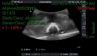
Today we scanned the pancreas, great vessels, liver, gall bladder and the commom bile duct in one hour. For the gall bladder, we had the patient in two different posotions: supine, and left lateral decubitus. The included image was obtained in the decubitus position, following the long axis of the gall bladder. I helps a lot to interrogate and obtained gall bladder images in two different positions, in case we need to asses for stones, polyps or sludge. Stones and sludge will be gravity dependent, while the polyps are not.
Thursday, October 7, 2010
DMS 110 lab 10-07-10 Abdomen US pictures
Today we scanned the pancreas ( for the first time), GV, and the liver all in one hour. I was able to complete all the required images, but not without help from my instructor, especially when looking for the pancreas. I found it harder to visualize, than all the other organs, since it does not have a capsule, and it varies from patient to patient. I can improve the speed of my study, over time, as I get more practice imaging a large number of people, and my eye gets used to recognizing where the pancreas lies.
Thursday, September 30, 2010
DMS 110 lab 09-30-2010
Today we scanned the biliary tree and we adjusted the brightnes gain, TCG's and the power knobs. I was able to find the CBD in the left decubitus position, and it measured 3.6 mm. My patient had a lot of gas, so I had to place her on the left side and use an intercostal window.
Thursday, September 23, 2010
DMS 110 lab liver and great vessels 09/23/2010
Today we scanned each other without using the book as the reference. I was able to obtain images in long and trans of the great vessels, and the liver. I measured the distal aorta both in long and trans, right before it bifurcates. My partner was a great vis for liver, mainly for portal veins, but a hard one for the celiac trunk in transverse.
Over all a great lab, I was able to remember all the required images needed for our protocol.
Over all a great lab, I was able to remember all the required images needed for our protocol.
Thursday, September 16, 2010
DMS 110 lab liver, kidneys, spleen, gallbladder, CBD
We scanner each other and looked for liver, gallbladder, CBD, both kidneys, spleen, aorta. We took measurements in both longitudinal, and transverse of the kidneys, GB, CBD, aorta, spleen. We also positioned the patient in a left decub for the GB.
Thursday, September 9, 2010
September 9th, 2010 DMS 110 lab
Today in lab we scanned each other, using the intercostal window. We looked at the liver, great vessels, spleen, right and left kidneys and the liver vasculature. We scanned in sagital, transverse, and coronal planes, when the patient was taking a deep breath, and when she was taking a shallow one. At the end of a deep breath in, we noticed that the liver and other organs moved inferiorly, and at the end of a shallow breath, the organs did not move as much.
The liver looked much clearer after the patient took a deep breath, because it moved inferiorly and was not superimposed by the ribs as much. The spleen looked better after a shallow inspiration, and the kidneys looked more clear when the patient was holding the breath.
The liver looked much clearer after the patient took a deep breath, because it moved inferiorly and was not superimposed by the ribs as much. The spleen looked better after a shallow inspiration, and the kidneys looked more clear when the patient was holding the breath.
Thursday, September 2, 2010
DMS 110 lab 09-01-2010
Today we scanned each other using the left hand, and paying attention to ergonomics. We scanned the aorta in longitudinal, but switching between the normal view and the harmonics view to see the difference.
Second DMS 120 lab 08-31-10
Today we scanned each other again, but we switched partners and machines. I learned to use the newest machine. The images were so much better.
First time we scanned each other
Today 08-26-10 in DMS 120 lab we scanned each other. We looked for the great vessels, both in longitudinal and in transverse cuts. The scanner that we used was not connected to Pacs, so I have no images to attach.
Tuesday, August 24, 2010
First DMS 110 lab
Today, we started our lab for DMS 110. We scanned bags filled with water, oranges, pickles, lego pieces, dental floss, rubber band, and cheese.
I used two machines for it: the old Siemens, and the small portable machine.
It was very useful, and I learned a lot about how to handle the probe, how to start the machines, etc.
I used two machines for it: the old Siemens, and the small portable machine.
It was very useful, and I learned a lot about how to handle the probe, how to start the machines, etc.
Monday, August 23, 2010
First day of school
Today is the first day of school, 8-23-2010. We started the day at 9:00 am with DMS 110-intro to Diagnostic Medical Sonography, followed by DMS 145-Clinical Pathology, at 10:00am. After a lunch break we had DMS 120-Abdominal Procedures.
Saturday, August 21, 2010
New student orientation
On Friday, August 20th, 2010 we had our new student orientation, from 8am to 12pm. It was filled with a lot of good information. I purchased my book package, which cost about $650.
The program will start on Monday, August the 23rd, at 9am.
I am excited, since I have been waiting for this for about 8 years.
The program will start on Monday, August the 23rd, at 9am.
I am excited, since I have been waiting for this for about 8 years.
Subscribe to:
Comments (Atom)


.bmp)
.bmp)
.bmp)
.bmp)


